View Featured Offers >>
77195
PathScan® RP BRD4 Sandwich ELISA Kit
ELISA Kits
ELISA Kit

PathScan® RP BRD4 Sandwich ELISA Kit #77195

Citations (0)
Figure 1. BRD4 protein levels in 293T cells are reduced upon transfection with siRNA directed against BRD4. The relationship between lysate protein concentration from 293T cells, either mock transfected or transfected with BRD4 siRNA, and the absorbance at 450 nm using the PathScan® RP BRD4 Sandwich ELISA Kit #77195 is shown in the upper figure. The corresponding western blots using BRD4 antibody (left panel) and α-Actinin antibody (right panel) are shown in the lower figure.
To Purchase # 77195
Cat. # Size Qty. Price
77195C
1 Kit  (96 assays)

Important Ordering Details

Custom Ordering Details:

When ordering five or more kits, please contact us for processing time and pricing.

Looking for this ELISA kit in a 384-well format? Inquire for availability, processing time, and pricing.

Supporting Data

REACTIVITY H M R Mk

Application Key:

  • WB-Western Blot
  • IP-Immunoprecipitation
  • IHC-Immunohistochemistry
  • ChIP-Chromatin Immunoprecipitation
  • C&R-CUT&RUN
  • C&T-CUT&Tag
  • DB-Dot Blot
  • eCLIP-eCLIP
  • IF-Immunofluorescence
  • F-Flow Cytometry

Species Cross-Reactivity Key:

  • H-Human
  • M-Mouse
  • R-Rat
  • Hm-Hamster
  • Mk-Monkey
  • Vir-Virus
  • Mi-Mink
  • C-Chicken
  • Dm-D. melanogaster
  • X-Xenopus
  • Z-Zebrafish
  • B-Bovine
  • Dg-Dog
  • Pg-Pig
  • Sc-S. cerevisiae
  • Ce-C. elegans
  • Hr-Horse
  • GP-Guinea Pig
  • Rab-Rabbit
  • All-All Species Expected
Product Includes Volume Solution Color
BRD4 Rabbit mAb Coated Microwells 96 tests
BRD4 Rabbit Detection mAb 1 ea Red (Lyophilized)
HRP Diluent 5.5 ml Red
TMB Substrate 7004 11 ml
STOP Solution 7002 11 ml
Sealing Tape 2 ea
ELISA Wash Buffer (20X) 9801 25 ml
Cell Lysis Buffer (10X) 9803 15 ml

Product Description

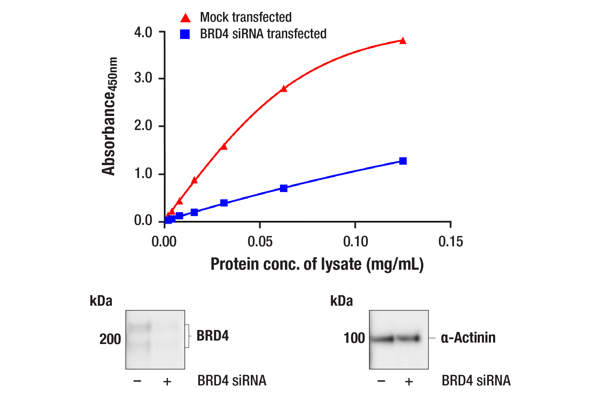
The rapid protocol (RP) PathScan® RP BRD4 Sandwich ELISA Kit is a solid phase sandwich enzyme-linked immunosorbent assay (ELISA) that detects endogenous levels of BRD4 protein in a reduced assay time of 1.5 hours. Incubation of cell lysates and detection antibody on the coated microwell plate forms a sandwich with BRD4 protein in a single step. The plate is then extensively washed and TMB reagent is added for signal development. The magnitude of absorbance for the developed color is proportional to the quantity of BRD4 protein. Learn more about your ELISA kit options here.

*Antibodies in this kit are custom formulations specific to kit.

Protocol

PRINT

View >Collapse >

PathScan® Sandwich ELISA Protocol (Rapid Protocol)

NOTE: This protocol is for PathScan® kits that use an HRP directly conjugated to the detection antibody (Rapid Protocol), rather than a 2-step method where the detection antibody and a secondary-HRP are added sequentially.

A. Solutions and Reagents

NOTE: Prepare solutions with deionized/purified water or equivalent.

  1. Microwell strips: Bring all to room temperature before opening bag/use. Unused microwell strips should be returned to the original re-sealable bag containing the desiccant pack and stored at 4°C.
  2. Detection Antibody: Reconstitute lyophilized Detection Antibody (red colored cake) with 1 mL of HRP Diluent (red solution) to yield a concentrated stock solution. Incubate at room temperature for 5 min with occasional gentle mixing to fully reconstitute. To make the final working solution, add the full 1 mL of reconstituted Detection Antibody to 4.5 mL of HRP Diluent in a clean tube and gently mix. For best results, use immediately following antibody reconstitution. Unused reconstituted Detection Antibody may be stored for up to 4 weeks at 4°C, although there may be some loss of signal compared to freshly reconstituted antibody.
  3. HRP Diluent: Red colored diluent for reconstitution and dilution of the Detection Antibody that is linked to HRP.
  4. 1X ELISA Wash Buffer: Prepare by diluting ELISA Wash Buffer (20X) (included in each kit) to 1X with deionized water.
  5. 1X Cell Lysis Buffer: Prepare by diluting 10X Cell Lysis Buffer #9803 to 1X with deionized water. This buffer can be stored at 4°C for short-term use (1–2 weeks). Recommended: When using to prepare cell lysates, add Protease/Phosphatase Inhibitor Cocktail (#5872, not supplied) and 1 mM phenylmethyl- sulfonyl fluoride (PMSF, #8553, not supplied) immediately before use.
  6. TMB Substrate (#7004): Bring to room temperature before use.
  7. STOP Solution (#7002): Bring to room temperature before use.

B. Preparing Cell Lysates

For adherent cells

  1. Aspirate media when the culture reaches 80–90% confluence. Treat cells by adding fresh media containing regulator for desired time.
  2. Remove media and rinse cells once with ice-cold 1X PBS.
  3. Remove PBS and add 0.5 mL ice-cold 1X Cell Lysis Buffer including 1 mM PMSF and Protease/Phosphatase Inhibitor Cocktail to each plate (10 cm diameter) and incubate the plate on ice for 5 min.
  4. Scrape cells off the plate and transfer to an appropriate tube. Keep on ice.
  5. Sonicate lysates on ice.
  6. Microcentrifuge for 10 min (14,000 rpm) at 4°C and transfer the supernatant to a new tube. The supernatant is the cell lysate. Store at −80°C in single-use aliquots.

For suspension cells

  1. Remove media by low speed centrifugation (~1200 rpm) when the culture reaches 0.5–1.0 x 10⁶ viable cells/mL. Treat cells by adding fresh media containing regulator for desired time.
  2. Collect cells by low speed centrifugation (~1200 rpm) and wash once with 5-10 mL ice-cold 1X PBS.
  3. Cells harvested from 50 mL of growth media can be lysed in 2.0 mL of 1X Cell Lysis Buffer including 1 mM PMSF and Protease/Phosphatase Inhibitor Cocktail.
  4. Sonicate lysates on ice.
  5. Microcentrifuge for 10 min (14,000 rpm) at 4°C and transfer the supernatant to a new tube. The supernatant is the cell lysate. Store at −80°C in single-use aliquots.

C. Test Procedure

NOTE: Equilibrate all materials and prepared reagents to room temperature prior to running the assay.

  1. Prepare all reagents as indicated above (Section A).
  2. Samples should be undiluted or diluted with 1X Cell Lysis Buffer to a 2X protein concentration in order to achieve a final 1X protein concentration upon addition of the Detection Antibody. Individual datasheets for each kit provide a sensitivity curve that serves as a reference for selection of an appropriate starting lysate concentration. The sensitivity curve shows typical results across a range of lysate concentration points.
  3. Add 50 µL of each sample to the appropriate wells.
  4. Add 50 µL of the Detection Antibody to each well.
  5. Seal the plate and incubate for 1 hour at room temperature on a plate shaker set to 400 rpm (moderate agitation).
  6. Gently remove the tape and wash wells:
    1. Discard plate contents into a receptacle.
    2. Wash 4 times with 1X Wash Buffer, 200 µL each time for each well.
    3. For each wash, strike plates on fresh towels hard enough to remove the residual solution in each well, but do not allow wells to completely dry at any time.
    4. Clean the underside of all wells with a lint-free tissue.
  7. Add 100 µL of TMB Substrate to each well. Seal with tape and incubate the plate in the dark for 15 min at room temperature on a plate shaker (400 rpm, moderate agitation) or alternatively for 10 min at 37°C without shaking.
  8. Add 100 µL of STOP Solution to each well. Shake gently for a few seconds.
  9. NOTE: Initial color of positive reaction is blue, which changes to yellow upon addition of STOP Solution.

  10. Read results:
    1. Visual Determination: Read within 30 min after adding STOP Solution.
    2. Spectrophotometric Determination: Wipe underside of wells with a lint-free tissue. Read absorbance at 450 nm within 30 min after adding STOP Solution.

created July 2020

Protocol Id: 2144

Specificity / Sensitivity

The PathScan® RP BRD4 Sandwich ELISA Kit detects endogenous levels of BRD4 protein. This kit recognizes the BRD4 long isoform (UniProt #O60885-1) and does not recognize other BRD4 isoforms. The kit sensitivity is shown in Figure 1. This kit detects proteins from the indicated species, as determined through in-house testing, but may also detect homologous proteins from other species.

Background

Bromodomain-containing protein 4 (BRD4) is a member of the bromodomains and extra terminal (BET) family of proteins, which also includes BRD2, BRD3, and BRDT (1-3). BET family proteins contain two tandem bromodomains and an extra terminal (ET) domain, and bind acetyl lysine residues (3). BRD4 is a chromatin-binding protein with a preference for Lys14 on histone H3 as well as Lys5 and Lys12 on histone H4 (4). BRD4 chromatin binding occurs throughout the cell cycle, including condensed mitotic chromosomes, when the majority of genes are silenced (5). BRD4 association with chromatin during mitosis is thought to be an important part of the bookmarking mechanism to accelerate re-activation of the silenced genes upon exit from mitosis (2,6). BRD4 has been shown to facilitate transcription by recruiting the positive transcription elongation factor b (pTEFb) complex that phosphorylates Ser2 of the heptapeptide repeat of the carboxy-terminal domain of RNA polymerase II, promoting transcription elongation (3,7,8). In addition, BRD4 has been found to be part of the super elongation complex and the polymerase associated factor complex (PAFc) in MLL-fusion derived leukemia cell lines, demonstrating a role for BRD4 in the regulation of transcription elongation (9). Research studies have shown that BRD4 (and BET family proteins) may be promising therapeutic targets for various Myc-driven cancers, such as Burkitt’s lymphoma and certain acute myeloid leukemias (1,10,11). Investigators have found molecular inhibition of BET proteins to be effective in inducing apoptosis in various MLL-fusion driven leukemic cell lines by competing BRD3 and BRD4 from chromatin, leading to reduced expression of Bcl-2, Myc, and CDK6 (9). BET inhibition has also been shown to have antitumor activities against nuclear protein in testis (NUT) midline carcinoma cell lines and xenografts in mice where BRD4 is found to be a frequent translocation partner of the NUT protein (12). In addition, BRD4 regulates the expression of some inflammatory genes, and inhibition of BRD4 (and BET family proteins) chromatin binding causes reduced expression of a subset of inflammatory genes in macrophages, leading to protection against endotoxic shock and sepsis (13).

Limited Uses

Except as otherwise expressly agreed in a writing signed by a legally authorized representative of CST, the following terms apply to Products provided by CST, its affiliates or its distributors. Any Customer's terms and conditions that are in addition to, or different from, those contained herein, unless separately accepted in writing by a legally authorized representative of CST, are rejected and are of no force or effect.

Products are labeled with For Research Use Only or a similar labeling statement and have not been approved, cleared, or licensed by the FDA or other regulatory foreign or domestic entity, for any purpose. Customer shall not use any Product for any diagnostic or therapeutic purpose, or otherwise in any manner that conflicts with its labeling statement. Products sold or licensed by CST are provided for Customer as the end-user and solely for research and development uses. Any use of Product for diagnostic, prophylactic or therapeutic purposes, or any purchase of Product for resale (alone or as a component) or other commercial purpose, requires a separate license from CST. Customer shall (a) not sell, license, loan, donate or otherwise transfer or make available any Product to any third party, whether alone or in combination with other materials, or use the Products to manufacture any commercial products, (b) not copy, modify, reverse engineer, decompile, disassemble or otherwise attempt to discover the underlying structure or technology of the Products, or use the Products for the purpose of developing any products or services that would compete with CST products or services, (c) not alter or remove from the Products any trademarks, trade names, logos, patent or copyright notices or markings, (d) use the Products solely in accordance with CST Product Terms of Sale and any applicable documentation, and (e) comply with any license, terms of service or similar agreement with respect to any third party products or services used by Customer in connection with the Products.

For Research Use Only. Not for Use in Diagnostic Procedures.
Cell Signaling Technology is a trademark of Cell Signaling Technology, Inc.
PathScan is a registered trademark of Cell Signaling Technology, Inc.
All other trademarks are the property of their respective owners. Visit our Trademark Information page.